4. Possible underlying neural mechanisms of BAVI
Pediatric BAVI as a neurodevelopmental disorder may affect the auditory and visual processing neural structures, pathways and underlying mechanisms. It might thus be conjectured that the spectrum of visual and auditory deficits seen in pediatric BAVI could be attributed to underlying brain dysfunction that affects the development of visual and auditory processing pathways and systems. However, it should be emphasized that the underlying relationships in BAVI are more complex and currently we cannot pinpoint exactly the level of neurological specificity.
The human brain is phenomenally complex and consists of regions and networks that exhibit a balance between network integration and segregation of visual and auditory processing. The brain is characterized by highly complex interactive networks. Current evidence suggests that the brain`s central associative hubs links regions within networks and function as an efficient “small world” connector that supports large-scale information integration (Li, et al., 2026).
Visual processing is the visual system’s ability to effectively transfer and analyze visual information, along the same lines auditory processing is the auditory system’s ability to effectively transfer and analyze auditory information. Visual and auditory processing are associated with a complex array of sensory impressions which are encoded respectively by the ears and eyes and then processed by multiple functional areas or pathways in the brain. The auditory and visual processing in the human brain suggest that the properties of sound (e.g., pitch, amplitude, timbre, duration) and the properties of vision (e.g., color, depth, motion, form) are processed in separate areas of the cerebral cortex and are assumed to be organized in parallel processing pathways (Salo, et al., 2013). Thus, efficient perceptual and cognitive functioning requires the integration of separate, but interconnected cortical networks in the brain (Parks, et. al., 2013).
According to the cortical processing of visual input, light or visual impressions from the environment are encoded by the retina which includes both the sensory neurons that respond to light and the intricate neural circuits that perform the first stages of image processing. The extracted aspects of the image are encoded as electrical messages, which travel through the optic nerve and optic chiasm. Signals are then transmitted along the thalamic neurons, conveyed through the optic chiasm before they reach the cerebral cortex for further feature identification and visual perception. The thalamus, specifically the lateral geniculate nucleus, actively relays visual information from the retina to the primary visual cortex in the occipital lobe for interpretation (Shipp, 2007). Visual areas beyond the primary visual cortex are called the extra striate visual cortex. The extra striate area V5/MT is crucial for processing the overall direction or coherent movement of a whole object or scene (Riecanský, 2004; Pamir et al., 2021). Furthermore, the neural regions of the extra striate cortex are considered part of either the ventral or dorsal stream (Milner, 2017). These pathways or networks are assumed to use visual object and visual spatial information differently (Salo, et al., 2013). The ventral network projects from the primary visual cortex to the inferior temporal cortex (i.e., occipitotemporal pathway). Generally, the ventral network (what” pathway), is associated with the encoding of basic geometric shapes, color discrimination and in the processing of foreground/background (level of depth) of visual stimuli. Information is then fed anteriorly to the inferotemporal cortex, where the fusiform face area resides, encoding facial features to enable efficient facial recognition (Prasad & Dinkin, 2019). In contrast, the dorsal network projects from the primary visual cortex to the posterior parietal cortex (i.e., occipitoparietal pathway). Largely, the dorsal network («where” pathway) is associated with motion processing and representations of object locations (Khader et. al., 2005). Besides, it is also responsible for processing the visual information needed for visually guided behavior (Norman, 2002). It is also crucial for visuospatial working memory, acting as the neural substrate for maintaining spatial information, location, and motion (Pisella, 2017). See figure 1.
According to the cortical processing of auditory input, sound or auditory impressions involve the collection of sound wave energy from the environment to the fluid vibrations within the cochlea. These sound vibrations or vibratory energy are then transformed into electric potentials by hair cells, and the activated neural impulses are transmitted via the auditory nerve. From the auditory nerve, auditory signals ascend along the central auditory pathways in which most critical sound attributes are processed. Signals are then transmitted along thalamic neurons before they reach the cerebral cortex. The thalamus, specifically the medial geniculate body, actively relays auditory information to the primary auditory cortex in the temporal lobe for interpretation (Lee, 2013). Auditory information enters the cortex through the primary auditory areas and then projects to secondary auditory areas. The primary auditory cortex is the first cortical region involved in acoustic processing, whereas the superior temporal gyrus encompasses the secondary auditory areas. The secondary auditory areas process more complex features, such as helps to organize sound information spatially (i.e. sound location) and interfaces with the lower-level auditory areas and higher-level structures contributing to language, music, and other forms of auditory processing (Ahmadi, et al., 2024).
Studies have demonstrated that non-auditory-related brain areas are critical for auditory processing (Schmithorst et al., 2013; Farah et al., 2014). For instance, the corpus callosum is involved in auditory processing because it is the main pathway for transferring auditory information between the brain's two hemispheres (Musiek, et al., 2011). Besides, the cerebellum, known to play an important role in motor functions, is also involved in a range of auditory processing functions such as coordinating timing and rhythm, processing temporal features of sound and in sound recognition through stored cerebellar memory templates (McLachlan & Wilson, 2017). Analogously to the visual “what-where” pathway model, two parallel processing streams (ventral/dorsal) have been identified in the auditory modality (Alain, et al., 2001). The ventral network projects from the primary auditory cortex ventrally along the temporal lobe to regions of the anterior temporal lobe, such as the anterior superior gyrus and Heschl's gyrus (Leavitt, et al., 2011). The ventral network ("what" pathway) is associated with processing the content, identity and meaning of sounds, and in the processing of short-timescale auditory patterns such as environmental and speech sounds (Ahveninen, et al., 2006). In contrast, the dorsal network projects from the primary auditory cortex dorsally to the regions of the parietal cortex, such as the planum temporal and posterior superior temporal gyrus (Leavitt, et al., 2011). Largely, the dorsal network (where” pathway) is associated with the spatial processing of sound, locating sound sources, processing of rapidly changing auditory information (Ahveninen, et al., 2006), and in maintaining and integrating sound-based items (Poliva, 2016). See figure 1.
As described, extending findings demonstrate a diverse range of independent neural systems that are engaged in visual and auditory processing. However, there are other neural systems that connect brain regions and form an anatomical basis for an integrated processing of visual and auditory information. For instance, distinct brain areas (i.e., posterior superior temporal sulcus & intraparietal sulcus) contribute to the integration of visual and auditory contents (Grefkes, & Fink, 2005; Beauchamp, et al., 2004).
It should also be emphasized that the functions of the ventral and dorsal networks are very much integrated and less segregated. A more nuanced network-level understanding of information processing, points towards the existence of more intricate context-driven functional networks selective of “what” and “where” information rather than segregated streams of processing along ventral and dorsal brain regions (Ray et al., 2020). Besides, the ventral and dorsal networks have distinct circuits but collaborative roles on attentional subprocesses. Neither of the two networks control attentional processes in isolation. The flexible interaction between both systems enables the dynamic control of attention in relation to bottom-up sensory stimulation and top-down goals (Vossel, et al., 2014). See fig. 1.
Bottom-up attention is important for detecting salient events, whereas top-down attention is important for maintaining continuous tracking of visual or auditory information (i.e., top-down selective attention). Selective attention is characterized by the tracking of visual or auditory objects while filtering out distracting visual or auditory disturbances.
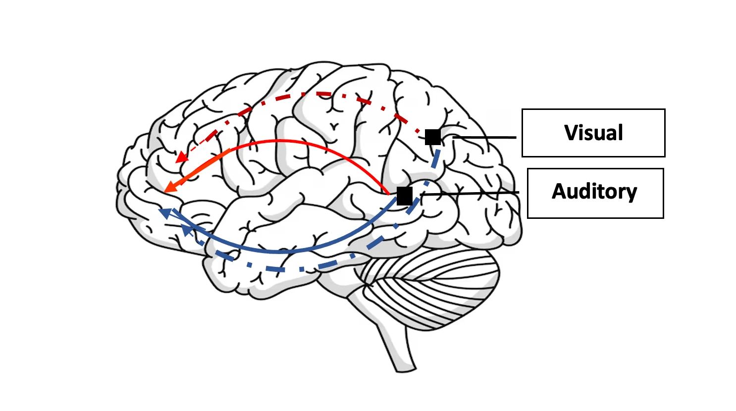
In terms of impairment, dysfunction in the visual brain areas or pathways may result in specific visual processing deficits. For instance, dysfunctions in the thalamus may disrupt the flow of visual input and result in visual perceptual processing deficits (Sherman, 2007), whereas interference in the V5/MT area might lead to a motion-specific visual discrimination deficit (Blanke, et al., 2002). Dysfunctions involving the ventral network are associated with difficulties with visual recognition and visual memory of objects, faces and other visual stimuli, whilst dysfunctions involving the dorsal network are associated with difficulties with visual attention, visuospatial relations, and with the visual guidance of movement (Binkofski & Buxbaum, 2013). Besides, dorsal network disruptions may result in difficulties of actively maintaining visual information (i.e., shapes, colors) and spatial information (i.e., location, movement) in memory over short periods of time (i.e., visuospatial working memory) (Pisella, 2017).
Likewise, dysfunctions in auditory brain areas or pathways may result in specific auditory processing deficits. For instance, dysfunction in the cerebellum might lead to significant disruptions in the perception of temporal aspects of sound and in difficulties processing the timing of sounds (Akshoomoff et al., 1994), whereas dysfunction in the corpus callosum may disrupt the flow of auditory input and result in auditory perceptual deficits (Chermak, et. al, 2011). Particularly, maldevelopment of the corpus callosum may interrupt auditory interhemispheric transfer resulting in greater difficulty localizing sound and understanding speech in noise (Bamiou, et al., 2007). Dysfunctions involving the ventral network are associated with difficulties in auditory object recognition, such as problems in identifying or recognizing sounds or vocal communications and problems in organizing sounds or speech into meaningful components, whilst dysfunctions involving the dorsal network are associated with difficulties with the spatial processing of sounds and difficulties in processing rapid auditory changes such as those in a conversation or in musical piece. Besides, dorsal network disruptions may result in difficulties temporarily storing and manipulating sound-based information and with difficulties of actively maintaining sounds in memory over short periods of time (i.e., auditory working memory) (Leavitt, et al., 2011; Poliva, 2016).
In general, dysfunctions of BAVI should be considered as breakdown in the whole brain's ability to process both auditory and visual information efficiently. Accordingly, one or more neural areas involved in auditory lower & higher order processes and visual lower order & higher order processes may be compromised or constrained in Brain based Visual and Auditory Impairment (BAVI). However, we lack evidence for the level of neural specificity of BAVI.
It is crucial to highlight that when exploring the dysfunctions of BAVI, neuroimaging can provide some valuable anatomical insights into the etiologies of BAVI. Neuroimaging techniques have significantly advanced our understanding of brain function and provided essential insights into neurological disorders (Yen, et al., 2023). For instance, a Magnetic Resonance Imaging (MRI) assessment would facilitate in revealing brain structures or brain networks and may aid in systematically understanding the underlying neural mechanisms associated with BAVI. However, it must be emphasized that BAVI may be present in a child, but cerebral damage or dysfunction per se may not always be detected by means of standard brain imaging methods. Importantly, auditory and visual processing problems may be linked to functional disruptions in specific auditory and visual pathways without demonstrability of structural brain changes. Thus, it is important to keep in mind that some children with BAVI may display functional deficits due to BAVI even when causative brain injury cannot be corroborated through structural brain imaging methods. This would suggest that, while structural brain abnormalities play a role in the etiology of BAVI, standard neuroimaging assessments (i.e., MRI) may be limited in their capability to correlate visual and auditory functional deficits with neuroanatomical changes. Whether the source of BAVI can or cannot be revealed by standard neuroimaging assessments or whether the impairment is mild or severe, the underlying source of BAVI is neurological, resulting from brain malfunctions or disruptions in brain functioning.